As You Know That I Have Colon Caner and I Might Not Able to Attend the Events
Colorectal cancer is extremely common. Symptoms include blood in the stool and change in bowel habits. Screening using one of several methods is recommended for appropriate populations. Diagnosis is by colonoscopy. Handling is surgical resection and chemotherapy for nodal interest.
-
1. Siegel RL, Miller KD, Jemal A: Cancer statistics, 2020. CA Cancer J Clin seventy(1):7–30, 2020. doi: x.3322/caac.21590
Patients in populations with a high incidence of CRC eat low-cobweb diets that are high in beast protein, fat, and refined carbohydrates. Carcinogens may exist ingested in the diet but are more than likely produced by bacterial action on dietary substances or biliary or abdominal secretions. The exact mechanism is unknown.
CRC spreads by direct extension through the bowel wall, hematogenous metastasis, regional lymph node metastasis, and perineural spread.
Symptoms and Signs of Colorectal Cancer
Colorectal adenocarcinomas grow slowly, and a long interval elapses earlier they are large enough to crusade symptoms. Symptoms depend on lesion location, blazon, extent, and complications.
The right colon has a large quotient and a thin wall and its contents are liquid; thus, obstruction is a late upshot. Bleeding is usually occult. Fatigue and weakness caused past astringent anemia may be the but complaints. Tumors sometimes grow large enough to exist palpable through the abdominal wall before other symptoms appear.
The left colon has a smaller lumen, the feces are semisolid, and cancer tends to crusade obstruction before than in the right colon. Partial obstruction with colicky intestinal hurting or consummate obstacle may exist the initial manifestation. The stool may exist streaked or mixed with blood. Some patients present with symptoms of perforation, commonly walled off (focal pain and tenderness), or rarely with diffuse peritonitis.
In rectal cancer, the most common initial symptom is bleeding with defecation. Whenever rectal bleeding occurs, even with obvious hemorrhoids or known diverticular disease, coexisting cancer must be ruled out. Tenesmus or a awareness of incomplete evacuation may be present. Pain is common with perirectal interest.
Some patients first present with symptoms and signs of metastatic illness (eg, hepatomegaly, ascites, supraclavicular lymph node enlargement).
-
Colonoscopy
-
Colonoscopy
-
Fecal occult blood testing
-
Sometimes flexible sigmoidoscopy
-
Sometimes fecal Deoxyribonucleic acid testing
-
Sometimes CT colonography
For average-risk patients, screening for colorectal cancer (CRC) should begin at age 45 years and go on until age 75 years. For adults anile 76 to 85, the decision whether to screen for CRC should exist individualized, taking into consideration the patient'due south overall wellness and prior screening history (see also the U.Due south. Preventive Services Chore Force's 2021 recommendation argument for screening for colorectal cancer and the American College of Gastroenterology's [ACG] clinical guidelines for colorectal cancer screening).
There are multiple options for CRC screening, including
-
Colonoscopy every ten years
-
Fecal occult blood test annually (fecal immunochemical tests [FIT] preferred)
-
Flexible sigmoidoscopy every 5 years (every 10 years if combined with FIT)
-
CT colonography every 5 years
-
Fecal DNA testing combined with FIT every 3 years
Fecal immunochemical tests for blood are more sensitive and specific for man blood than older guaiac-based stool tests, which tin be affected by many dietary substances. However, a positive test for blood can result from nonmalignant disorders (eg, ulcers, diverticulosis), and a negative test does not dominion out cancer because cancers practise not bleed continuously.
Fecal Dna testing detects Dna mutations and methylation markers shed from a colonic tumor. The test typically is combined with FIT, and the combined exam is approved for screening average-risk patients. Patients with a positive fecal Dna-FIT test should go a follow-up colonoscopy inside 6 months to reduce the hazard of missing advanced colon cancer. About ten% of patients with a positive fecal Deoxyribonucleic acid-FIT examination result have a normal colonoscopy; such patients can accept a repeat fecal DNA-FIT exam in one year or a repeat colonoscopy in 3 years. If these tests are negative, they can return to the average-chance colon cancer screening schedule.
Video capsule endoscopy of the colon has many technical problems and is non currently acceptable as a screening test.
Claret-based tests (eg, Septin 9 assay) accept been approved for screening boilerplate-risk patients merely are non widely used because of inadequate sensitivity.
-
Colonoscopic biopsy
-
CT to evaluate extent of tumor growth and spread
-
Genetic testing
Patients with positive fecal occult blood tests crave colonoscopy, as do those with lesions seen during sigmoidoscopy or an imaging written report. All lesions should exist completely removed for histologic exam. If a lesion is sessile or not removable at colonoscopy, surgical excision should be strongly considered.
Barium enema x-ray, specially a double-contrast study, can detect many lesions just is somewhat less accurate than colonoscopy and is not currently acceptable as follow-up to a positive fecal occult blood test.
Once cancer is diagnosed, patients should accept abdominal CT, chest x-ray, and routine laboratory tests to seek metastatic disease and anemia and to evaluate overall condition.
Elevated serum carcinoembryonic antigen (CEA) levels are present in seventy% of patients with CRC, but this exam is neither sensitive nor specific and therefore is not recommended for screening. However, if the CEA level is high preoperatively and low after removal of a colon tumor, monitoring the level may assist notice recurrence earlier. CA 19-ix and CA 125 are other tumor markers that may be similarly used.
Staging Colorectal Cancer*
| Stage | Tumor (Maximum Penetration) | Regional Lymph Node Metastasis | Distant Metastasis |
|---|---|---|---|
| 0 | Tis | N0 | M0 |
| I | T1 or T2 | N0 | M0 |
| Ii | T3 | N0 | M0 |
| III | Any T or | Whatsoever N | M0 |
| T4 | N0 | M0 | |
| IV | Whatever T | Any Northward | M1 |
| * TNM classification:
| |||
-
Surgical resection, sometimes combined with chemotherapy, radiation, or both
Surgery for cure tin can be attempted in the lxx% of patients presenting without metastatic affliction. Endeavour to cure consists of wide resection of the tumor and its regional lymphatic drainage with reanastomosis of bowel segments.
For rectal cancer, sphincter-saving surgical resection can be washed in patients with rectal cancer that has a distal margin of ≥ 1.0 cm, instead of the usual five-cm length, without significant hazard of local recurrence or decreased long-term survival. Sphincter-saving procedures have been done in patients with rectal cancer that has a distal margin of < 1cm, but these patients take an increased hazard of local recurrence and decreased long-term survival. The problem with sphincter-saving procedures is often more functional (eg, fecal leakage, incontinence) in nature rather than oncologic (eg, local recurrence, decreased survival). If there is local recurrence or poorly tolerated bowel function after a sphincter-saving procedure, then an abdominoperineal resection (April) with permanent colostomy (1 Treatment references Colorectal cancer is extremely mutual. Symptoms include blood in the stool and modify in bowel habits. Screening using one of several methods is recommended for appropriate populations. Diagnosis... read more than 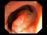 ) is done.
With liver metastases, resection of a limited number (1 to 3) of liver metastases is recommended in select nondebilitated patients as a subsequent procedure. Criteria include patients whose master tumor has been resected, whose liver metastases are in one hepatic lobe, and who have no extrahepatic metastases. Simply a small number of patients with liver metastases meet these criteria, but in such cases five-year postoperative survival is 25%.
Chemotherapy improves survival by at least 10 to xxx% in colon cancer patients with positive lymph nodes. Rectal cancer patients with 1 to 4 positive lymph nodes benefit from combined radiation and chemotherapy; when > iv positive lymph nodes are found, combined modalities are less constructive. Preoperative radiation therapy and chemotherapy to meliorate the resectability rate of rectal cancer or decrease the incidence of lymph node metastasis are standard.
Additional screening for recurrence should include history, concrete examination, and serum carcinoembryonic antigen levels every 3 months for iii years so every half dozen months for 2 years. Imaging studies (CT or MRI) are often recommended at 1-year intervals only are of uncertain benefit for routine follow-upwardly in the absence of abnormalities on examination or claret tests.
When curative surgery is not possible or the patient is an unacceptable surgical risk, limited palliative surgery (eg, to salve obstruction or resect a perforated area) may exist indicated; median survival is 7 months. Some obstructing tumors can be debulked past electrocoagulation or held open past stents. Chemotherapy may compress tumors and prolong life for several months.
Newer drugs used singly or in drug combinations include capecitabine (a v-fluorouracil forerunner), irinotecan, and oxaliplatin. Monoclonal antibodies such every bit bevacizumab, cetuximab, and panitumumab are also being used with some effectiveness. No regimen is clearly more than constructive for prolonging life in patients with metastatic colorectal cancer, although some have been shown to filibuster disease progression. Chemotherapy for advanced colon cancer should exist managed by an experienced chemotherapist who has admission to investigational drugs.
When metastases are confined to the liver simply cannot be surgically resected, hepatic artery infusion with floxuridine or radioactive microspheres, given either intermittently in a radiology department or given continuously via an implantable subcutaneous pump or an external pump worn on the chugalug, may offer more do good than systemic chemotherapy; still, these therapies are of uncertain do good. When metastases are likewise extrahepatic, intrahepatic arterial chemotherapy offers no advantage over systemic chemotherapy. For selected patients with ≤ three liver lesions, stereotactic radiations therapy or thermal ablation using radiofrequency or microwave treatments tin be considered.
-
1. Bujko K, Rutkowski A, Chang GJ, et al: Is the 1-cm rule of distal bowel resection margin in rectal cancer based on clinical testify? A systematic review. Ann Surg Oncol nineteen(iii):801–808, 2012. doi: 10.1245/s10434-011-2035-2
-
2. Kahi CJ, Boland R, Dominitz JA, et al: Colonoscopy surveillance afterward colorectal cancer resection: Recommendations of the U.s. multi-society task strength on colorectal cancer. Gastroenterology 150:758–768, 2016. doi: ten.1053/j.gastro.2016.01.001
-
Colorectal cancer is 1 of the nigh mutual cancers in western countries, typically arising inside an adenomatous polyp.
-
Right-sided lesions usually manifest with haemorrhage and anemia; left-sided lesions usually manifest with obstructive symptoms (eg, colicky abdominal pain).
-
Routine screening should brainstorm at age 45 for patients with average risk; typical methods involve colonoscopy or annual fecal occult blood testing and/or flexible sigmoidoscopy.
-
Serum carcinoembryonic antigen (CEA) levels are often elevated but are not specific enough to exist used for screening; however, afterward handling, monitoring CEA levels may assistance detect recurrence.
-
Treatment is with surgical resection, sometimes combined with chemotherapy and/or radiation; outcome varies widely depending on the stage of the disease.
The following are some English-linguistic communication resources that may be useful. Please note that THE Transmission is not responsible for the content of these resources.
Source: https://www.msdmanuals.com/professional/gastrointestinal-disorders/tumors-of-the-gastrointestinal-tract/colorectal-cancer
0 Response to "As You Know That I Have Colon Caner and I Might Not Able to Attend the Events"
Post a Comment